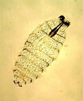
We reported the case of a 21-year-old female patient from Sakarya, Turkey, who visited the local hospital with pain in the right costo-lumbar region. She also complained of irritation in the vagina, dysuria, polyuria, hematuria, vomiting and diarrhea. During her stay in the hospital five grayish-brown larvae, approximately 3 mm in length, were isolated from her urine. The larvae were identified as the fourth stage of the moth fly Psychoda albipennis (Taylan-Ozkan et al. 2004).
The case of an eight-year-old boy with hyper-eosinophilia who presented with a swelling under his right armpit that had persisted for more than four weeks, was reported. A second-stage larva of Wohlfahrtia magnifica was found, leading to the diagnosis of cutaneous myiasis. Following removal of the larva, the clinical and hematological manifestations returned to normal. Diagnosis of myiasis should always be kept in mind in the event of clinical signs of furuncular lesions, which may be accompanied by eosinophilia (Tuygun et al. 2009).
A 33 year old woman living in Elad, a small city about 25 km east of Tel Aviv in the middle of a rural area, had suffered for years from sinusitis and asthma for which she was treated with antibiotics and prednisone. Recently she was not permitted to take part in nature trips and had not left the country for the last 2 years. On 20 May 2010, she observed two small living larvae that were expelled from her nose while sneezing. The larvae were sent to a neighboring hospital but were lost. On 2 October 2010, a third, larger, larva was discharged from her nose while sneezing, and was brought to our laboratories for examination. The larva was black in color and had shrunk, indicating that it had been dead for some time before it was expelled from the nasal cavity. It was identified as the third stage of Oestrus ovis (Mumcuoglu & Ron, 2010).
A retrospective observational study of patients who presented with myiasis was conducted between 1999 and July 2014 in the post-travel clinics in Israel. Data regarding exposure history, travel duration, clinical presentation, treatment, and parasitological identification were collected and analyzed. Among 6,867 ill returning Israeli travelers, 1,419 (21%) had a dermatologic complaint, 90 (6.3%) of them were diagnosed with myiasis. Myiasis was acquired in Latin America by 72 (80%) patients, mainly (54%) in the Madidi National Park, Amazonas Basin, Bolivia; 18 cases (20%) were acquired in Africa. Of the 18 African cases, 15 were identified as infested by C. anthropophaga, while all the infestations caused by C. rodhaini were acquired in Ghana, while all cases from South America were diagnosed as Dermatobia hominis. In 76% of cases, manual extraction was sufficient to remove the larva; 24% required surgical intervention. Despite the fact that most patients did not receive antibiotic treatment, only one developed secondary infection, upon partial removal of the larva (Lachish et al. 2015).
We described the case of a post traumatic myiasis due to Wohlfahrtia magnifica in a mixed Pit-Bull Terrier dog in Israel. The dog was successfully treated by removing the maggots, administering a larvicidal endectocide, ivermectin, and treating the wound with antibiotics and antiseptic topical washes (Salant et al. 2020).
References:
Ayalon A, Yehezkeli V, Paitan Y, Szpila K, Mumcuoglu KY, Moisseiev E. 2020. Massive orbital myiasis caused by Sarcophaga argyrostoma complicating eyelid malignancy. Case Rep Ophthalmol Med. 2020 Apr 30;2020:5618924. doi: 10.1155/2020/5618924.
Lachish T, Marhoom E, Mumcuoglu KY, Tandlich M, Schwartz E. Myiasis in travelers. 2015. J. Travel Med. doi: 10.1111/jtm.12203.
Mumcuoglu, K.Y. & E. Ron. 2011. Nasal myiasis due to Oestrus ovis larvae in Israel. Israel Med. Assoc. J. 13: 379-380.
Salant H, Mumcuoglu KY, Baneth G. 2020. Myiasis in a dog in Jerusalem. Isr J Vet Med. 75(4): 221-224
Taylan-Ozkan, A., C. Bahur, S. Kilic, S. Nalbantoglu, I. Dalkilic & K.Y. Mumcuoglu. 2004. Urogenital myiasis caused by Psychoda albipennis (Diptera: Nematocera) in Turkey. Int. J. Dermatol. 43: 904-905.
Tuygun, N., A. Taylan-Ozkan, G. Tanir & K.Y. Mumcuoglu. 2009. Furuncular myiasis in a child caused by Wohlfahrtia magnifica (Diptera: Sarcoptidae) associated with eosinophilia. Turkish J. Pediatr. 51: 275-281.
Additional publication on this subject:
Mumcuoglu, K.Y. and T. Rufli. 1980. Dermatological entomology. 11. Myasis (in German). Schweiz. Rundschau Med. 69:912-922.
Mumcuoglu, K.Y. 2014. Other Ectoparasites: Leeches, Myiasis and Sand Flies. In: Manson’s Tropical Diseases. Farrar, J., Hotez, P.J., Junghanss, T., Kang, G., Lalloo, D. & N.J. White (Eds.), 23th. Edition, Elsevier, pp. 843-847.
