Research Projects
Defining the axonal trajectories of spinal IN and their neuronal circuit.
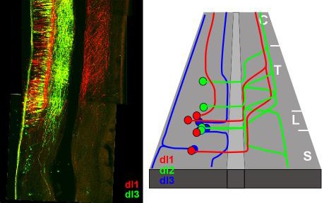
Employment of neuronal subtype enhancer elements to drive expression of reporter genes in neurons is a widely used paradigm for deciphering neuronal circuits. Several caveats hamper the use of enhancer elements for tracking axonal trajectories of spinal interneurons (INs): a) There are few genes that are expressed uniquely in specific IN subpopulations; instead, a combinatorial code of gene expression determines the neuronal fate of INs. b) Only few IN enhancer elements have been characterized. c) For tracking axonal projection of INs in vertebrates, germ line-targeted reporter genes yield bilaterally-symmetric labeling. Therefore, it is hard to distinguish between the ipsi- and contra-laterally projecting axons. Utilizing transient-transgenic chick screening of highly conserved non-coding sequences, and employing enhancer intersection techniques, we have identified numerous enhancer elements that are expressed in spinal neuronal subpopulations. To this end, tools for tracking the axonal projection and targeting of the dorsal interneuron populations: dI1, dI2 and dI3, were generated. We found that each dI neuronal population has a unique axonal patterning that differs in the ipsi/contra-lateral, rostral/caudal and the ventral/dorsal axes, and specific neuronal targets (Avraham et al., 2010a; Avraham et al., 2009; Avraham et al., 2010b; Mansour et al., 2010). Thus, laying the foundation for studying the mechanism of circuit formation in the neural tube.
Elucidating the transcriptional code that governs the axonal patterning of INs.
A transcriptional code distinguishes dI1-3 neurons. The expression of the Lim-homeodomain (Lim-HD) proteins Lhx2/9, Lhx1/5 and Isl1 is restricted to dI1, dI2 and dI3 neurons, respectively. To begin to understand the possible transcriptional control of Lim-HD on dI1-3 axonal projection, the Lim- HD code of dI neurons was altered by cell-specific gain and loss of function. Our data point to an instructive role for Lim-HD proteins in patterning the axonal trajectories of dI1-3 neurons. Lhx9, Lhx1 and Isl1 are sufficient to confer dI1, dI2 and dI3 axonal patterning, respectively (Avraham et al., 2010a; Avraham et al., 2009).
Determining the role of guidance molecules at axonal choice points
Axons change their trajectory plane at precise choice points. The floor plate, the ventral midline cells of the spinal cord, is a choice point for axons turning either ipsi or contra-laterally and caudally or rostrally. The floor plate is also a transition point from axons extending in a cellular milieu (gray matter) to elongation at the basement membrane that ensheathes the neurons (the white matter). F-spondin is a guidance molecule expressed at the floor plate. The in vivo proteolysis of F-spondin generates two functionally opposing fragments: a short-range repellent protein deposited in the membrane of floor plate cells and an adhesive protein that accumulates at the basement membrane. Their coordinated activity, acting respectively as a short-range repellent and a permissive short-range attractant, constricts commissural axons to the basement membrane beneath the floor plate cells. Thus, proteolysis coordinates combinatorial guidance signaling originating from a single guidance cue. Our studies reveal that an additional level of complexity in guidance cues resides by modification of guidance molecules. Similar posttranslational modifications may augment the versatility of other guidance molecules and thus intensify the diversity of guidance cues (Feinstein and Klar, 2004; Zisman et al., 2007).

Characterization of neuronal receptors for positional guidance cues.
Previous studies have implicated the midline of the spinal cord – the roof plate and the floor plate, as the sole source of guidance molecules in the spinal cord. Our studies have revealed novel axonal routes and en-passant intermediate guidance cues for interneurons - the axons of motor and sensory neurons (Avraham et al., 2010a). Our current and future studies are aimed toward deciphering the molecular codes that instruct the axonal projection along these cues.
References
Avraham, O., Hadas, Y., Vald, L., Hong, S., Song, M.R., and Klar, A. (2010a). Motor and Dorsal Root Ganglion Axons Serve as Choice Points for the Ipsilateral Turning of dI3 Axons. J Neurosci 30, 15546-15557.
Avraham, O., Hadas, Y., Vald, L., Zisman, S., Schejter, A., Visel, A., and Klar, A. (2009). Transcriptional control of axonal guidance and sorting in dorsal interneurons by the Lim-HD proteins Lhx9 and Lhx1. Neural Dev 4, 21.
Avraham, O., Zisman, S., Hadas, Y., Vald, L., and Klar, A. (2010b). Deciphering axonal pathways of genetically defined groups of neurons in the chick neural tube utilizing in ovo electroporation. J Vis Exp.
Feinstein, Y., and Klar, A. (2004). The neuronal class 2 TSR proteins F-spondin and Mindin: a small family with divergent biological activities. The Inter J of Bioch & Cell Biol 36, 975–980.
Mansour, A.A., Nissim-Eliraz, E., Zisman, S., Golan-Lev, T., Schatz, O., Klar, A., and Ben-Arie, N. (2010). Foxa2 regulates the expression of Nato3 in the floor plate by a novel evolutionarily conserved promoter. Mol Cell Neurosci in press.
Zisman, S., Marom, K., Avraham, O., Rinsky-Halivni, L., Gai, U., Kligun, G., Tzarfaty-Majar, V., Suzuki, T., and Klar, A. (2007). Proteolysis and membrane capture of F-spondin generates combinatorial guidance cues from a single molecule. J Cell Biol 178, 1237-1249.
