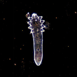
 Demodex folliculorum
 and
Demodex brevis are obligatory parasites in hair follicles and in pilosebaceous glands of human skin. Although most people are infested with these mites, only a small number develop the clinical symptoms of skin demodicosis.
Twenty-five patients with human demodicosis and 150 controls were typed for HLA-A, B, Bw, and Cw using the microlymphocytotoxicity method. The immune response was evaluated by identifying membrane markers of different immune cells using monoclonal antibodies. An association between the frequency of HLA Cw2 and Cw4 haplotypes and human demodicosis was established. The risk of developing clinical symptoms of this disease is 5.0 times higher for people with the Cw2 phenotype and 3.1 times higher for those with the Cw4 haplotype. Individuals who have the HLA A2 phenotype are 2.9 times more resistant to demodicosis. A positive correlation between demodicosis and the haplotypes A3-Cw4, A3-Cw2, A3-B17, A3-B35 and B35-Cw4 was found. In addition, an association between Cw2 and Cw4 alleles in the phenotype of patients with demodicosis and a decrease in the number of NK cells was found (Akilov & Mumcuoglu, 2003).
Twenty-nine patients with human demodicosis and thirteen age-and-sex-matched healthy subjects participated in the study. The presence of mites was determined by microscopic inspection of secretion from sebum glands. The immune response was evaluated in the peripheral blood by identifying membrane markers of different immune cells using monoclonal antibodies, while the concentration of IgA, IgM, and IgG was calculated by simple radial immunodiffusion using anti-IgA, IgM, and IgG. The level of circulating immune complexes and total haemolytic complement, as well as the preparatory and digestive function of neutrophils, and the functional activity of leukocytes were also studied. The absolute numbers of CD95+ was higher in patients with demodicosis. The absolute number of CD3+, CD4+, CD8+, and CD16+ cells, the ratio of CD3+/CD20+, and the functional activity of leukocytes were significantly lower in individuals infested with Demodex mites. No significant differences were found in the percentage and absolute number of CD20+ cells, the ratio of CD4+/CD8+ T cell subpopulations, circulating immune complexes, CH50, activity and index of phagocytosis and the level of IgM, IgG and IgA antibodies between individuals infested with Demodex mites and the control group. It is concluded that the readiness of lymphocytes to apoptosis increases parallel to increasing density of mites. This could be the result of a local immunosuppression caused by mites, which allows them to survive in the host skin (Akilov & Mumcuoglu, 2004).
The goal of this study was to study the correlation between immunological and immunogenetic data obtained from patients with demodicosis in order to clarify the pathogenesis of Demodex infestation. Twenty-five patients with demodicosis and 13 age- and sex-matched healthy subjects participated in the study. The presence of mites was determined by microscopic inspection of sebum gland secretions. The immune response was evaluated by identifying membrane markers of different immune cells using monoclonal antibodies (anti-CD3+, CD4+, CD8+, CD16+, CD20+ and CD95+) while the concentration of IgA, IgM and IgG was measured by simple radial immunodiffusion. The level of circulating immune complexes and total hemolytic complement as well as the preparatory and digestive function of neutrophils and the functional activity of leukocytes were also studied. Patients were typed for HLA A, B, Bw and Cw using the microlymphocytotoxicity method. The comparison between patients with and without the A2 phenotype showed that the latter have lower numbers of CD8+, lower functional activity of leukocytes, higher concentrations of IgA, larger affected skin areas and are more often affected by deep papular and papulo-pustular forms of demodicosis than those with the A2 phenotype, showing that this allele has a protective role in demodicosis. Patients exhibiting the Cw2 phenotypes were rather susceptible to demodicosis. They showed decreased numbers of CD3+, increased levels of phagocytic activity, higher mite density and severer skin damage as compared to patients lacking Cw2. It is argued that HLA A2 and Cw2 phenotypes have an important diagnostic, prognostic and pathogenetic significance and could play a role in resistance or susceptibility to demodicosis by regulating the end phase of the immune response (Mumcuoglu & Akilov, 2005).
To characterize the clinical features and course of the different forms of demodicosis, a comparative, observational, clinical, acarological and longitudinal study was conducted. Eighty-seven patients with clinical symptoms of demodicosis and positive acarological findings during the period December 1999 to December 2001 were examined. The usual etiological agent of primary demodicosis is
D. folliculorum, which causes an erythematic squamous skin eruption in the central part of the face. The efflorescence appears on unaltered skin and covers 11.8 ± 3.8% of the face. The accompanying pruritus appears together with the efflorescence. The erythema appears after the papulo-pustules is due to inflammation and disappears after treatment. In 51.2% of the patients, no seasonal exacerbation of demodicosis can be seen. In addition to skin alteration, there are concomitant gastrointestinal and urological complications. The secondary demodicosis is mainly caused by
D. brevis and it characterized by a papulo-pustular eruption in the middle of the face. The efflorescence appears on an altered skin due to a dermatosis of different origin and covers 35.2±4.9% of the face. The pruritus starts after the appearance of the efflorescence. The erythema appears before the papulo-pustular eruption, is due to vessel dystonia and continues after the treatment. In most patients it becomes worse during the hot months of the year. In addition, cardiovascular, gynecological, gastrointestinal and endocrine ailments are observed in these patients. It is concluded that for the differentiation of the different forms of demodicosis, in addition to the form of dermatosis, their distribution, seasonality, pathogenicity as well as the species of mite involved must be taken into consideration (Akilov et al. 2005).
The follicle mites
Demodex folliculorum and
Demodex brevis could play a role in several skin and ocular diseases, however in lower numbers these mites could also be found in healthy individuals. Demodicosis is a chronic skin disease, which affects mainly the face and it is caused by the presence of large number of follicle mites. Rosacea complicated by Demodex mite infestation and Demodex blepharitis present therapeutic difficulties in everyday practice. Acaricidal formulations containing lindane, crotamiton, permethrin, sulfur, mercury and ivermectin have been shown to be effective in diminishing the number of mites and alleviating the symptoms of demodicosis (Mumcuoglu & Akilov, 2010).
The aim of this study was to determine the prevalence and density of Demodex folliculorum and Demodex brevis in students and staff of the Erzincan University, Turkey, and to define the influence of age, gender, educational level, and hygiene as well as skin moisture, pH, and temperature on the presence of Demodex mites. Healthy people without apparent facial dermatoses from nine faculties and five vocational schools of the university were included in the study. The measurements for moisture, pH, and temperature were conducted in the cheek region, while samples for mite presence were taken from the same region using the standard superficial skin biopsy technique. A total of 538 healthy people, 385 students and 153 university staff, were included in the study. Demodex mites were detected in 50.1% of the students (mean 7.1/cm2 ) and in 69.3% of the university staff (mean 13.1/cm2 ). There were statistical differences between the groups of people who were cleaning their face one, two, three, or more times daily and between those who were using and not using personal towels. There was a borderline significant difference between the different age groups and people with different education levels. The density of mites was higher in those with skin moisture of less than 50%, with a pH of 5-6.5 and temperature of 24-28 °C, however the differences between the groups with different skin moisture, skin pH, and skin temperature were not statistically significant (Zeytun et al. 2017).
The aim of this study was to determine the prevalence and Demodex density in sickle cell anemia (SCA) patients and to compare with healthy subjects. The study included 70 patients diagnosed with SCA and control group of 50 healthy individuals. Samples were taken from cheeks, forehead, nose, and chin and were examined microscopically. Infestation of ≥5 mites/cm2 was accepted as positive in the diagnosis. Demodex mite positivity was determined in 20 (28.6%) patients and none in subjects of the control group. In the SCA group, the mean number of mites was 26.10/cm2. A statistically significant correlation was found between Demodex mite positivity and the number of SCA symptom attacks experienced by the patients within the last 1 yr (P ≤ 0.001). No significant relationship was determined between Demodex mite positivity and age or gender (P = 0.56 and P = 0.11, respectively). Demodex mites are seen more often in SCA patients who suffer from a compromised immune system, and the presence of Demodex mites could be a risk factor in the appearance of SCA symptom attacks (Kaya et al. 2019).
In this study, our aim was to investigate the skin-homing T-cell immune responses triggered in patients with Demodex infestation and/or rosacea. Collected whole blood samples were divided into four groups: control subjects; non-rosacea patients with Demodex infestation (Demodex group); papulopustular rosacea (PPR) patients without Demodex infestation (Rosacea group); and PPR patients with Demodex infestation (Rosacea/Demodex group). Following ex vivo activation, skin-homing CLA+CD4+ T-cell subset levels were monitored by flow cytometry. When compared with control subjects, among skin-homing CD4+ T-cell subsets analyzed, Demodex patients had higher TH 9 and Treg cell levels; Rosacea subjects displayed elevated TH 1 cell levels; and Rosacea/Demodex patients exhibited increased frequencies of TH 9 and TH 22 cells. In contrast to Rosacea subjects, Rosacea/Demodex group members displayed higher TH 2 cell levels; and when compared with Demodex groups, they had higher TH 1 and TH 2 but lower Treg cell levels. Demodex group members also exhibited higher Treg but lower TH 1 and TH 22 levels than Rosacea/Demodex group subjects (Gazi et al. 2019a).
The aim of this review was to summarize the relevant literature on demodicosis obtained from studies conducted on humans and dogs, and draw the attention to the effect of mite-associated factors (e.g., microbiota) on the different clinical manifestations displayed during human and canine demodicosis. Canine and human demodicosis are caused by different Demodex species, and the clinical manifestations in former could be life-threatening. Nevertheless, current literature suggests similar immune responses and immune evasion mechanisms in human and canine demodicosis; cellular immunity appeared to have a central role in protection against demodicosis, and Demodex mites were shown to influence both innate and adaptive immune response to escape immune attack (Gazi et al. 2019b).
References
Akilov OE, Mumcuoglu KY. 2003. Association between human demodicosis and HLA class I. Clin. Exp. Dermatol. 28: 70-73.
Akilov OE, Mumcuoglu KY. 2004. Immune response in demodicosis. J. Eur. Acad. Dermatol. Venereol. 18: 440-444.
Akilov OE, Butov YS, Mumcuoglu KY. 2005. A clinico-pathological approach to the classification of human demodicosis. J. German Soc. Dermatol. 3:607-614.
Gazi U, Gureser AS, Oztekin A, Karasartova D, Kosar-Acar N, Derici MK, Artuz F, Mumcuoglu KY, Taylan-Ozkan A. 2019a. Skin-homing T-cell responses associated with Demodex infestation and rosacea. Parasite Immunol. 24:e12658. doi: 10.1111/pim.12658.
Gazi U, Taylan-Ozkan A, Mumcuoglu KY. 2019b. Immune mechanisms in human and canine demodicosis: A review. Parasite Immunol. 2019 Sep 26:e12673. doi:10.1111/pim.12673.
Kaya OA, Akkucuk S, Ilhan G, Guneri CO, Mumcuoglu K. 2019. The importance of Demodex mites (Acari: Demodicidae) in patients with sickle cell anemia. J. Med. Entomol. 56(3): 599-602. doi: 10.1093/jme/tjy225.
Mumcuoglu KY, Akilov OE. 2005. The role of HLA A2 and CW2 in the pathogenesis of human demodicosis. Dermatology 210: 109-114.
Mumcuoglu KY, Akilov OE. 2010. The role of Demodex mites in the pathogenesis of rosacea and blepharitis and their control. J. Rosacea Res. Dev. Inst. 1: 47-54.
Zeytun E, Tilki E, Doğan S, Mumcuoğlu KY. 2017. The effect of skin moisture, pH, and temperature on the density of Demodex folliculorum and Demodex brevis (Acari: Demodicidae) in students and staff of the Erzincan University, Turkey. Int J Dermatol. 56(7): 762-766. doi:10.1111/ijd.13600.
Additional publications on this subject
Mumcuoglu, K.Y. 1985. Demodex infestation of goats in Switzerland. In: Acarology VI. Griffiths, D.A. and C.E. Bowman (eds). Vol. II, Horwood, Chichester, p. 1132-1137.
Rufli, T. and K.Y. Mumcuoglu. 1981. The hair follicle mites Demodex folliculorum and D. brevis: Biology and importance in human medicine. Dermatologica 162:1-11.
Rufli, T., K.Y. Mumcuoglu, A. Cajacob and S. Buchner. 1981. Demodex folliculorum: The ethiopathogenis and therapy of rosacea and perioral dermatitis (in German). Dermatologica 162:12-26.
Rufli, T., K.Y. Mumcuoglu, A. Cajacob and S. Buchner. 1981. Dermatological entomology. 22. Demodicidae/Follicle mites (in German). Schweiz. Rundschau Med. 70:622-630.
