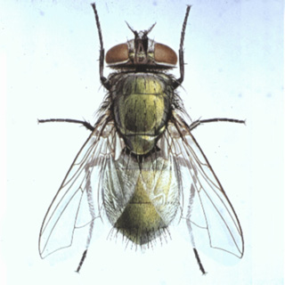
The beneficial effect of fly larvae for wounds was first observed by Ambroise Pare, in the 16th Century. Baron Larrey, physician-in-chief to Napoleon's armies and Dr. Joseph Jones, a medical officer during the American Civil war, while working with soldiers who remained wounded for several days in the battle fields, described that maggots destroy only dead tissue, and do not injure living tissue.
Maggot debridement therapy (MDT), the treatment of suppurative skin infections with the larvae of calliphorid flies, was first introduced by Baer in 1931. This method was used extensively in the 1930's and early 1940's in over 300 hospitals in the USA alone, and was abandoned later with the introduction of antibiotics and the use of aggressive surgical debridement. Since then maggots were used only occasionally as salvage therapy for skin and soft tissue wounds which did not respond to surgery and antibiotic therapy. Since 1989 in the USA and since mid-1990's in Great Britain Germany, Sweden, Switzerland and Israel MDT has been re-introduced for the treatment of intractable wounds. During the last 20 years over 80,000 patients have been treated by this method (Mumcuoglu, 2001).
Clinical work
Since its introduction to Israel in 1996, the Maggot Debridement Therapy (MDT) has been used in over 3,000 patients with ca. 4,800 wounds; 600 of them was treated by myself, while the remaining with maggots, which were sent from my laboratory or the CWT company to over 20 hospitals and clinics in Israel. A full debridement was achieved in 81% of the cases, while by over 100 patients, an imminent leg amputation could be prevented. Sterile maggots (24-48 hrs old) of the green bottle fly,
Lucilia sericata were administered to the wound 2-5 times weekly and replaced every 1-3 days. Depending on the size of the wound the number of treatments varied between 1-8 (median: 2, average 3), and lasted for a period of 1-12 days (median: 3, average: 4.7). As the therapy progressed, new layers of healthy tissue were formed over the wounds, while the offensive odor emanating from the necrotic tissue and the intense pain accompanying the wound decreased significantly. The majority of the patients did not complain about any major discomfort during the treatment. In ca. 30% of the patient the existing pain increased as a result of MDT.
It was observed that maggots are capable of entering any part of the wound wherever necrotic tissue exists and clean minute areas without harming healthy tissue in a manner resembling micro-surgery; a task which is very difficult to attain by the conventional surgery. After MDT, patients were sent either for a skin transplant or their wounds were treated with hydrocolloidal sheets or disinfectants.
Maggot therapy is a rapid and effective treatment alternative, indicated for large necrotic wounds requiring debridement when conventional treatment and conservative surgical intervention do not help (Gilead et al. 2012, Mumcuoglu et al., 1997, 1999, 2001, 2012; Sherman et al. 2011).
This treatment modality was also introduced by the present author to Turkey (three centers), as well as in Switzerland and Tanzania. MDT was accepted as a medical device in May 2011 by the Israeli Ministry of Health and was introduce to the "Sal Habriut". Between 2012 and 2019 disinfected maggots were produced and distributed by the CWT company.
Antibacterial substances from maggots
Green fluorescent protein-producing Escherichia coli were used to investigate the fate of bacteria in the alimentary tract of sterile grown maggots,
Lucilia sericata (Meigen), using a laser scanning confocal microscope. A computer program was used to analyze the intensity of the fluorescence and to quantify the number of bacteria. The crop and the anterior midgut were the most heavily infected areas of the intestine. A significant decrease in the amount of bacteria was observed in the posterior midgut. The number of bacteria decreased even more significantly in the anterior hindgut and practically no bacteria were seen in the posterior end, near the anus. The viability of bacteria in the different gut sections was examined. It was shown that 66.7% of the crops, 52.8% of the midguts, 55.6% of the anterior hindguts, and 17.8% of posterior hindguts harbored living bacteria. In conclusion, during their passage through the digestive tract the majority of E. coli was destroyed in the midgut. Most of the remaining bacteria were killed in the hindgut, indicating that the feces were either sterile or contained only small numbers of bacteria (Mumcuoglu et al. 2001).
Extracts of sterile and non-sterile maggots showed an activity of 200 arbitrary units (AU)/ml and 400AU/ml respectively. Maggots removed from chronic wounds had an activity of 1200AU/ml. Injuring sterile maggots with a sterile needle doubled the antibacterial activity within 24 hours, while the antibacterial activity of hemolymph increased fourfold after injury with a sterile needle and sixteen-fold with an infected needle. The fractions with a molecular weight of < 1kDa and 3-10kDa showed antibacterial activity against Gram-positive and Gram-negative bacteria including Pseudomonas aeruginosa, Klebsiella pneumoniae and methicillin-resistant Staphylococcus aureus (MRSA) isolated from wounds. The fraction with a molecular weight of < 1kDa lysed over 90% of the bacteria within 15 minutes by causing an influx of K+ and changing the membrane potential of bacteria. It was concluded that the nature of the antibacterial materials extracted from maggots not only indicates their ability to ingest the necrotic tissue on the wound, but also their potential significance in wound healing (Huberman et al. 2007a).
To partially characterize maggot-secreted antibacterial substances and determine their range of activity against different bacteria, sterile and non-sterile maggots maintained in the laboratory and taken from chronic wounds of treated patients were used. Whole body extracts and hemolymph were fractionated and their range of activity against bacteria was tested using the zone of inhibition assay. The mode of action of bacterial destruction was examined by viable counts, influx of K+ and changes in the membrane potential by scanning electron microscope. Low molecular weight compounds were isolated by high-performance liquid chromatography from the maggot or hemolymph extracts of
Lucilia sericata. Using gas chromatography-mass spectrometry analysis, three compounds were obtained: p-hydroxybenzoic acid (molecular weight 138 Da), p-hydroxyphenylacetic acid (molecular weight 152 Da) and octahydro-dipyrrolo[1,2-a;1',2'-d] pyrazine-5,10-dione (molecular weight 194 Da), also known as the cyclic dimer of proline (or proline diketopiperazine or cyclo[Pro,Pro]). All three molecules revealed antibacterial activity when tested against Micrococcus luteus and/or Pseudomonas aeruginosa, and the effect was even more pronounced when these molecules were tested in combination and caused lysis of these bacteria (Huberman et al. 2007b).
Maggots of the green blowfly,
Lucilia sericata, are used as an alternative to surgical intervention and long-term antiseptic therapy for the treatment of chronic wounds. The secretions of maggots are known to have antibacterial properties. To quantify the bactericidal effect of secretions from larvae of
L. sericata, an in vitro test model based on the modified European quantitative suspension test (EN 1040) was developed, in which a co-culture of maggots and bacteria (Micrococcus luteus, Escherichia coli, methicillin-sensitive Staphylococcus aureus) in tryptic soy broth was tested. The numbers of bacterial colonies with and without maggot exposure were compared after 24, 48 and 72 h of exposure. The mean log 10 reduction factor (RF) for bacterial elimination per maggot was 1 4 at all examined times for all tested bacteria. Thus, maggot secretion fulfilled the required definitions of an antiseptic. In addition, the maggots' ability to ingest bacteria was also evaluated. Maggots contained viable bacteria after 48 h of contact with the respective organisms. These maggots also continued excreting bacteria. Therefore, maggots should be disposed of after use as they must be regarded as medical waste (Daeschlein et al. 2007).
During 1996–2009, 723 wounds of 435 patients (180 females and 255 males) were treated with maggot debridement therapy (MDT) in 16 departments and units of the Hadassah Hospital in Jerusalem, Israel. Overall, 261 patients were treated during hospitalization, while 174 were treated as ambulatory patients. In 90.5% of the patients the wounds were located on the leg, but only 48.0% had diabetic foot ulcers. The wound duration range from one to 240 months (mean=8.9; median=4 months). Sterile maggots of the green bottle fly,
Lucilia sericata, were used for MDT. In 90.6% of the cases, maggots were placed directly on the wound using a cage-like dressing and left for 24 hours, while in 9.4% of the patients maggots concealed in a tea-bag like polyvinyl netting were used. The concealed maggots were left on the wound for 2–3 days. The number of treatments was 1–48 (mean=2.98; median=2) and the duration of the treatment varied between one and 81 days (mean=4.65; median=3). In 357 patients (82.1%) complete debridement of the wound was achieved, while in 73 patients (16.8%) the debridement was partial and in five (1.1%) it was ineffective. Increased pain or discomfort during MDT were reported in 38% of the patients (Gilead et al. 2012).
Chronic wounds are still regarded as a serious public health concern, which are on the increase mainly due to the changes in life styles and aging of the human population. Maggot therapy (MT) was initially thought to act mainly through debridement, today MT is known to influence all four overlapping physiological phases of wound repair: homeostasis, inflammation, proliferation, and remodeling/maturing. During MT, medical-grade larvae are applied either freely or enclosed in tea-bag like devices (biobag) inside the wounds, which suggests that larva excretion/secretion (ES) products can facilitate the healing processes directly without the need of direct contact with the larvae. This review summarizes the relevant literature on ES-mediated effects on the cellular responses involved in wound healing (Gazi et al. 2021).
Review articles
Gilead et al. 2012; Sherman et al. 2013, 2021; Mumcuoglu & Taylan-Ozkan, 2021; Mumcuoglu, 2022, 2023; Tekin et al. 2022; Colak et al. 2023.
Webpage
KYM is the webmaster of the “International Biotherapy Society”
websiteCase report
The patient, a 75-year-old male, had been suffering from lymphostasis for about 50 years, and was hospitalized in a clinic for the chronically ill in Jerusalem. Two months earlier, after receiving a small injury, the patient developed sepsis and renal failure, and gangrene developed on his left leg below the knee. Serological tests indicated that he was infected with a strain of Streptococcus A. After surgical debridement and disinfection three times daily, the wound became heavily infected (Fig. 1). The patient suffered intense pain and it was impossible to transplant skin onto the leg at this stage and amputation of the leg was recommended.
Approximately 1,000 maggots, 48 hrs old, were placed on the wound daily for 5 days a week, and left for 24 hrs before being replaced by new larvae. The maggots cleaned the entire infected area and healthy granulation appeared after two weeks of treatment (Fig. 2). Meanwhile the pain diminished significantly. Thereafter the patient was referred for autologous skin transplantation.
Figure 1. Before treatment
Figure 2: After treatment
References
Daeschlein, G., K.Y. Mumcuoglu, O. Assadian, B. Hoffmeister & A. Kramer. 2007. In vitro antibacterial activity of Lucilia sericata maggot secretions. Skin Pharmacol. Physiol. 20:112–115 (DOI: 10.1159/000097983).
Gazi U, Taylan-Ozkan A, Mumcuoglu KY. 2021. The effect of Lucilia sericata excretion/secretion (ES) products on cellular responses in wound healing. Med Vet Entomol. 35: 257–266. doi: 10.1111/mve.12497.
Gilead, L., K.Y. Mumcuoglu & A. Ingber. 2012. The use of maggot debridement therapy in the treatment of chronic wounds in hospitalised and ambulatory patients. J. Wound Care 21: 78-85.
Huberman, L., N. Gollop, K.Y. Mumcuoglu, C. Block & R. Galun. 2007a. Antibacterial properties of the whole body extracts and hemolymph of Lucilia sericata maggots. J. Wound Care 16: 123-127.
Huberman, L., N. Gollop, K.Y. Mumcuoglu, E. Breuer, S.R. Bhusare, Y. Shai & R. Galun. 2007b. Antibacterial substances of low molecular weight isolated from Lucilia sericata (Diptera: Muscidae). Med. Vet. Entomol. 21: 127-131.
Mumcuoglu, K.Y. 2001. Clinical applications for maggots in wound care. Am. J. Clin. Dermatol. 2: 219-227.
Mumcuoglu, K.Y., M. Lipo, I. Ioffe-Uspensky, J. Miller, I. Turkeltaub & R. Galun. 1997. Maggot therapy for the treatment of gangrene and osteomyelitis. Harefuah 132: 323-325.
Mumcuoglu, K.Y., A. Ingber, L. Gilead, J. Stessman, R. Friedman, H. Schulman, H. Bichucher, I. Ioffe-Uspensky, J. Miller, R. Galun & I. Raz. 1998. Maggot therapy for the treatment of diabetic foot ulcers. Diabetes Care, 21: 2030-2031.
Mumcuoglu, K.Y., A. Ingber, L. Gilead, J. Stessman, R. Friedmann, H. Schulman, H. Bichucher, I. Ioffe-Uspensky, J. Miller, R. Galun & I. Raz. 1999. Maggot therapy for the treatment of intractable wounds. Intnl. J. Dermatol. 8: 623-627.
Mumcuoglu, K.Y., J. Miller, M. Mumcuoglu, M. Friger & M. Tarshis. 2001. Destruction of bacteria in the digestive tract of the maggot of Lucilia sericata (Diptera: Calliphoridae). J. Med. Entomol. 38: 161-166.
Mumcuoglu, K.Y., E. Davidson, A. Avidan & L. Gilead. 2012. Pain related to maggot debridement therapy. J. Wound Care 21: 400-405.
Sherman, R. A., J. M-T Sherman, L. Gilead, M. Lipo & K.Y. Mumcuoglu. 2001. Maggot debridement therapy in outpatients. Arch. Phys. Med. Rehabil. 82: 1226-1229.
Additional publications on this subject
Brin, Y.S., K.Y. Mumcuoglu, S. Massarwe, M. Wigelman, E. Gross & M. Nyska. 2007. Chronic foot ulcer management using maggot debridement and topical negative pressure therapy. J. Wound Care 16:111-113.
Çolak B, Taylan Özkan A, Mumcuoğlu KY. 2023. Traditional methods used in wound treatment: Larval debridement therapy and hirudotherapy (in Turkish). In: Yastı AÇ, Akın M (eds.). The wound. Akademisyen Kitabevi A.Ş., Ankara, Turkey, pp. 311-328.
Gazi U, Taylan Özkan A, Mumcuoglu KY. 2019. Larval therapy and chronic wounds (in Turkish). J Biotechnol Strategic Health Res. 3: 55-60. DOI: bshr.536577.
Mumcuoglu KY. 2015. Biotherapy in Israel (in Turkish). Tanyuksel, M. & K.Y. Mumcuoglu (Eds). Multidisciplinary and Biologically Based Natural Therapies – Biotherapy (Apitherapy, Hirudotherapy, Maggot Therapy and Ichthiotherapy) (in Turkish). Meta Basım, İzmir.
Mumcuoğlu KY. 2022. Application of Larval Debridement Therapy (in Turkish). In: Tekin A, Doğruman Al F, Mumcuoğlu KY (eds). Larval Debridement Therapy. Turkish Ministry of Health Publication No: 1236, ISBN: 978-975-590-846-5, Chapter 6.
LinkMumcuoglu, K.Y. & L. Gilead. 2013. Debridement therapy with medicinal maggots for the treatment of intractable wounds in Israel (in Hebrew). J. Isr. ABC Healing Problem Wounds, June 2013, pp. 27-30.
Mumcuoglu K.Y. & A. Taylan-Ozkan. 2009. The treatment of suppurative chronic wounds with maggot debridement therapy (in Turkish). Türkiye Parazitol. Derg. 33: 307-315.
Mumcuoglu KY & Taylan-Ozkan A. 2015. Maggot therapy: Worldwide (in Turkish). Tanyuksel, M. & K.Y. Mumcuoglu (Eds). Multidisciplinary and Biologically Based Natural Therapies – Biotherapy (Apitherapy, Hirudotherapy, Maggot Therapy and Ichthiotherapy) (in Turkish). Meta Basım, İzmir.
Mumcuoglu KY, Taylan-Ozkan A. 2021. The use of maggot therapy in the treatment of diabetic foot ulcers (in Turkish). Proceedings of the First International Kocaeli Traditional and Complementary Medicine Congress. June 11-13, 2021, Kocaeli, Turkey, pp. 112-122.
LinkMumcuoglu KY, Colak B, Taylan-Ozkan A. 2023. Maggot therapy for the treatment of decubitus ulcers (in Turkish). Anadolu Tibbi Dergisi 2(1): 22-28.
Razumov, S., I. Avraham, N. Porat, A. Ingber & K.Y. Mumcuoglu. 2009. Treatment of intractable wounds with maggots (in Hebrew). HaAhot BeIsrael XX: 15-19.
Sherman, R.A., K.Y. Mumcuoglu, M. Grasberger & T.I. Tantawi. 2013. Maggot Therapy. In: Grassberger, M., R.A. Sherman, O. Gileva, C.M.H. Kim & K. Y. Mumcuoglu (Eds). Biotherapy - History, Principles and Practice: A Practical Guide to the Diagnosis and Treatment of Disease using Living Organisms. Springer Pub., Heidelberg, pp. 5-29.
Sherman RA, Chon R. (Competency Committee: Armstrong DG, Bernard L, Bingham PA, Dorsey R, Fonseca-Munoz A, Lavery LA, Mendez S, Mumcuoglu KY, Niezgoda J, Rogers LC, Contreras-Ruiz J, Tantawi TI, Tippett A, Vincent C). 2021. BioTherapeutics, Education and Research Foundation position paper: Assessing the competency of clinicians performing maggot therapy. Wound Rep Reg. 2021;1-7. doi:10.1111/wrr.12986.
Tanyuksel, M., E. Araz, K. Dundar, G. Uzun, T. Gumus, B. Alten, F. Saylam, A. Taylan-Ozkan & K.Y. Mumcuoglu. 2005. Maggot debridement therapy in the treatment of chronic wounds in a military hospital setup in Turkey. Dermatology 210: 115-118.
Tanyuksel, M. & K.Y. Mumcuoglu (Eds). 2015. Multidisciplinary and Biologically Based Natural Therapies – Biotherapy (Apitherapy, Hirudotherapy, Maggot Therapy and Ichthiotherapy). Multidisipliner Yaklaşımlı Biyolojık Temelli Doğal Tedaviler – Biyoterapi (Apiterapi, Hirudoterapi, Maggot tedavi ve İhtiyoterapi) (in Turkish). Meta Basım, İzmir.
Taylan-Ozkan, A. & K.Y. Mumcuoglu. 2007. Maggot debridement therapy for the treatment of a venous stasis ulcer (in Turkish). Turkish Bull. Hyg. Exp. Biol. 64: 31-34.
Tekin A, Doğruman Al F, Mumcuoğlu KY (eds). 2022. Larval Debridement Therapy (in Turkish). Turkish Ministry of Health Publication No: 1236, ISBN: 978-975-590-846-5.
Link 
